Tag Archives: negative

Uridine Triphosphate Inhibits Bone Growth in Rat Cells In Vitro

Abstract

Osteoblast responses to nucleotides increase during differentiation.

Accumulating evidence suggests that extracellular nucleotides, signaling through P2 receptors, play a role in modulating bone cell function. ATP and ADP stimulate osteoclastic resorption, while ATP and UTP are powerful inhibitors of bone formation by osteoblasts. We investigated changes in the expression of P2 receptors with cell differentiation in primary osteoblast cultures. Rat calvarial osteoblasts, cultured for up to 10 days, were loaded with the intracellular Ca(2+)-sensing fluorophore, Fluo-4 AM, and a fluorescence imaging plate reader was used to measure responses to nucleotide agonists. Peak responses occurred within 20 s and were evoked by ATP or UTP at concentrations as low as 2 microM. Osteoblast number doubled between day 4 and 10 of culture, but the peak intracellular Ca(2+) response to ATP or UTP increased up to 6-fold over the same period, indicating that osteoblast responsiveness to nucleotides increases as cell differentiation proceeds. The approximate order of potency for the most active nucleotide agonists at day 8 of culture was ATP > UTP and ATPgammaS > ADP > UDP, consistent with the expression of functional P2Y(2), P2X(2), P2Y(4), P2Y(1) and P2Y(6) receptors. Smaller responses were elicited by 2-MeSATP, Bz-ATP and alpha,beta-meATP, additionally suggesting the presence of functional P2X(1), P2X(3), P2X(5) and P2X(7) receptors. Expression of mRNA for the ATP- and UTP-selective P2Y(2) receptor increased strongly between day 6 and 15 in primary rat osteoblasts, whereas mRNAs for the P2Y(4) (also ATP/UTP selective) and P2Y(6) (UDP/UTP selective) receptors were highly expressed at intermediate time points. In contrast, mRNA for the cell-proliferation-associated P2X(5) receptor decreased to undetectable as osteoblasts matured, but mRNA for the cell-death-associated P2X(7) receptor was detected at all time points. Similar trends were evident using immunostaining and Western blotting for P2 receptors. Exposure to 10 muM ATP or UTP during days 10-14 of culture was sufficient to cause near-total blockade of the ‘trabecular’ bone nodules formed by osteoblasts; however, UDP and ADP were without effect. Our results show that there is a shift from P2X to P2Y expression during differentiation in culture, with mature osteoblasts preferentially expressing the P2Y(2) receptor and to a lesser extent P2Y(4) and P2Y(6) receptors. Taken together, these data suggest that the P2Y(2) receptor, and possibly the P2Y(4) receptor, could function as ‘off-switches’ for mineralized bone formation.

Orriss IR, Knight GE, Ranasinghe S, Burnstock G…
Bone Aug 2006
PMID: 16616882


Abstract

ATP and UTP at low concentrations strongly inhibit bone formation by osteoblasts: a novel role for the P2Y2 receptor in bone remodeling.

There is increasing evidence that extracellular nucleotides act on bone cells via multiple P2 receptors. The naturally-occurring ligand ATP is a potent agonist at all receptor subtypes, whereas ADP and UTP only act at specific receptor subtypes. We have reported that the formation and resorptive activity of rodent osteoclasts are stimulated powerfully by both extracellular ATP and its first degradation product, ADP, the latter acting at nanomolar concentrations, probably via the P2Y1 receptor subtype. In the present study, we investigated the actions of ATP, ADP, adenosine, and UTP on osteoblastic function. In 16-21 day cultures of primary rat calvarial osteoblasts, ADP and the selective P2Y1 agonist 2-methylthioADP were without effect on bone nodule formation at concentrations between 1 and 125 microM, as was adenosine. However, UTP, a P2Y2 and P2Y4 receptor agonist, known to be without effect on osteoclast function, strongly inhibited bone nodule formation at concentrations >or= 1 microM. ATP was inhibitory at >or= 10 microM. Rat osteoblasts express P2Y2, but not P2Y4 receptor mRNA, as determined by in situ hybridization. Thus, the low-dose effects of extracellular nucleotides on bone formation and bone resorption appear to be mediated via different P2Y receptor subtypes: ADP, signalling through the P2Y1 receptor on both osteoclasts and osteoblasts, is a powerful stimulator of osteoclast formation and activity, whereas UTP, signalling via the P2Y2 receptor on osteoblasts, blocks bone formation by osteoblasts. ATP, the ‘universal’ agonist, can simultaneously stimulate resorption and inhibit bone formation. These findings suggest that extracellular nucleotides could function locally as important negative modulators of bone metabolism, perhaps contributing to bone loss in a number of pathological states.

Hoebertz A, Mahendran S, Burnstock G, Arnett TR
J. Cell. Biochem. 2002
PMID: 12210747


Abstract

Regulation of the osteogenic and adipogenic differentiation of bone marrow-derived stromal cells by extracellular uridine triphosphate: The role of P2Y2 receptor and ERK1/2 signaling.

An imbalance in the osteogenesis and adipogenesis of bone marrow-derived stromal cells (BMSCs) is a crucial pathological factor in the development of osteoporosis. Growing evidence suggests that extracellular nucleotide signaling involving the P2 receptors plays a significant role in bone metabolism. The aim of the present study was to investigate the effects of uridine triphosphate (UTP) on the osteogenic and adipogenic differentiation of BMSCs, and to elucidate the underlying mechanisms. The differentiation of the BMSCs was determined by measuring the mRNA and protein expression levels of osteogenic- and adipogenic-related markers, alkaline phosphatase (ALP) staining, alizarin red staining and Oil Red O staining. The effects of UTP on BMSC differentiation were assayed using selective P2Y receptor antagonists, small interfering RNA (siRNA) and an intracellular signaling inhibitor. The incubation of the BMSCs with UTP resulted in a dose-dependent decrease in osteogenesis and an increase in adipogenesis, without affecting cell proliferation. Significantly, siRNA targeting the P2Y2 receptor prevented the effects of UTP, whereas the P2Y6 receptor antagonist (MRS2578) and siRNA targeting the P2Y4 receptor had little effect. The activation of P2Y receptors by UTP transduced to the extracellular signal-regulated kinase 1/2 (ERK1/2) signaling pathway. This transduction was prevented by the mitogen-activated protein kinase inhibitor (U0126) and siRNA targeting the P2Y2 receptor. U0126 prevented the effects of UTP on osteogenic- and adipogenic-related gene expression after 24 h of culture, as opposed to 3 to 7 days of culture. Thus, our data suggest that UTP suppresses the osteogenic and enhances the adipogenic differentiation of BMSCs by activating the P2Y2 receptor. The ERK1/2 signaling pathway mediates the early stages of this process.

Li W, Wei S, Liu C, Song M…
Int. J. Mol. Med. Jan 2016
PMID: 26531757 | Free Full Text

High Salt Diet May be a Risk Factor for Osteoporosis in Korean Women

Abstract

Association between Urinary Sodium Excretion and Bone Health in Male and Female Adults.

High salt intake is a well-known risk factor for osteoporosis, but the association between bone mass and urinary sodium excretion has not been studied as yet. This study investigates the hypothesis that urinary sodium excretion is negatively associated with bone mass and the risk of osteoporosis.
This cross-sectional study was performed using data from the Korea National Health and Nutrition Examination Survey, 2008-2011. Participants (n = 16,279) were divided into age groups; men were categorized as younger than 50 years of age or 50 years or greater, women were categorized as pre- or post-menopausal. Multivariate linear regression analysis showed that urinary sodium excretion was negatively associated with bone mineral content (BMC) and bone mineral density (BMD) in premenopausal and postmenopausal women. Sodium excretion was negatively associated with BMC and BMD of the lumbar spine in women with normal bone health, osteopenia and osteoporosis, but there was no association in men. Increased sodium excretion was significantly associated with risk for osteoporosis/osteopenia in premenopausal women. This study demonstrates that urinary sodium excretion is negatively associated with bone health, suggesting that high salt intake could be a possible risk factor for osteoporosis in Korean women, but not in men.

Park Y, Kwon SJ, Ha YC
Ann. Nutr. Metab. 2016
PMID: 26967579

Can Leaky Gut Cause Bone Loss?

Abstract

Epithelial Barrier Function in Gut-Bone Signaling.

The intestinal epithelial barrier plays an essential role in maintaining host homeostasis. The barrier regulates nutrient absorption as well as prevents the invasion of pathogenic bacteria in the host. It is composed of epithelial cells, tight junctions, and a mucus layer. Several factors, such as cytokines, diet, and diseases, can affect this barrier. These factors have been shown to increase intestinal permeability, inflammation, and translocation of pathogenic bacteria. In addition, dysregulation of the epithelial barrier can result in inflammatory diseases such as inflammatory bowel disease. Our lab and others have also shown that barrier disruption can have systemic effects including bone loss. In this chapter, we will discuss the current literature to understand the link between intestinal barrier and bone. We will discuss how inflammation, aging, dysbiosis, and metabolic diseases can affect intestinal barrier-bone link. In addition, we will highlight the current suggested mechanism between intestinal barrier and bone.

Rios-Arce ND, Collins FL, Schepper JD, Steury MD…
Adv. Exp. Med. Biol. 2017
PMID: 29101655

Green Tea Extract is Bad for Growing Bones in Rats

Abstract

Long-Term Intake of Green Tea Extract Causes Mal-Conformation of Trabecular Bone Microarchitecture in Growing Rats.

The purpose of this study was to examine the effects of green tea extract (GTE) intake on bone structural and physiological properties, such as bone mass, trabecular bone microarchitecture, cortical bone geometry, and bone mechanical strength, in growing rats. Four-week-old male Wistar rats were divided into the following four eenoups: standard diet feeding for 85 days (S-CON) or 170 days (L-CON), and GTE diet feeding for 85 days (S-GTE) or 170 days (L-GTE). At the end of the experiment, in addition to measurement of circulating bone formation/resorption markers, bone mass, trabecular bone microarchitecture, and cortical bone geometry were analyzed in the left femur, and bone mechanical strength of the right femur was measured. There was no difference in all bone parameters between the S-CON and S-GTE groups. On the other hand, the L-GTE group showed the decrease in some trabecular bone mass/microarchitecture parameters and no change in cortical bone mass/geometry parameters compared with the L-CON group, and consequently the reduction in bone weight corrected by body weight. There was no difference in bone formation/resorption markers and bone mechanical strength between the S-CON and S-GTE groups and also between the L-CON and L-GTE groups. However, serum leptin levels were significantly lower in the L-GTE group than in the L-CON group. Thus, the long-term GTE intake had negative effects on bone, especially trabecular bone loss and microarchitecture mal-conformation, in growing rats.

Minematsu A, Nishii Y, Imagita H, Sakata S
Calcif. Tissue Int. Nov 2017
PMID: 29103160

Milk Increases Risk of Fracture in Women

Abstract

Milk, dietary calcium, and bone fractures in women: a 12-year prospective study.

This study examined whether higher intakes of milk and other calcium-rich foods during adult years can reduce the risk of osteoporotic fractures.
This was a 12-year prospective study among 77761 women, aged 34 through 59 years in 1980, who had never used calcium supplements. Dietary intake was assessed with a food-frequency questionnaire in 1980, 1984, and 1986. Fractures of the proximal femur (n = 133) and distal radius (n = 1046) from low or moderate trauma were self-reported on biennial questionnaires.
We found no evidence that higher intakes of milk or calcium from food sources reduce fracture incidence. Women who drank two or more glasses of milk per day had relative risks of 1.45 for hip fracture (95% confidence interval [CI] = 0.87, 2.43) and 1.05 for forearm fracture (95% CI = 0.88, 1.25) when compared with women consuming one glass or less per week. Likewise, higher intakes of total dietary calcium or calcium from dairy foods were not associated with decreased risk of hip or forearm fracture.
These data do not support the hypothesis that higher consumption of milk or other food sources of calcium by adult women protects against hip or forearm fractures.

Feskanich D, Willett WC, Stampfer MJ, Colditz GA
Am J Public Health Jun 1997
PMID: 9224182 | Free Full Text

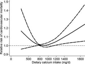
Calcium More or Less Than 800mg Increases Heart Risk, More or Less Than 900mg Increases All-Cause Mortality

Abstract

Dietary calcium intake and mortality risk from cardiovascular disease and all causes: a meta-analysis of prospective cohort studies.

Considerable controversy exists regarding the association between dietary calcium intake and risk of mortality from cardiovascular disease and all causes. Therefore, we performed a meta-analysis of prospective cohort studies to examine the controversy.
We identified relevant studies by searching MEDLINE, Embase, and the Cochrane Library databases between 1 September 2013 and 30 December 2013. Reference lists of relevant articles were also reviewed. Observational prospective studies that reported relative risks and 95% confidence intervals for the association of calcium intake with cardiovascular and all-cause mortality were eligible. Study-specific relative risks were pooled using a random-effects model.
In this meta-analysis, 11 prospective studies with 12 independent cohorts, involving 757,304 participants, were eligible. There was evidence of a non-linear association between dietary calcium intake and risk of mortality from cardiovascular disease (P for non-linearity <0.01) and all causes (P for non-linearity <0.01). A dose-response analysis showed a U-shaped relationship between dietary calcium intake and cardiovascular mortality. Intakes that were lower and higher than around 800 mg/day were gradually associated with a higher risk of cardiovascular mortality. For all-cause mortality, we also observed a threshold effect at intakes around 900 mg/day. The risk of all-cause mortality did not decrease further at intakes above 900 mg/day.
This meta-analysis of prospective cohort studies suggests that dietary calcium intake is associated with cardiovascular mortality in a U-shaped manner and that high dietary calcium intake (>900 mg/day) is not associated with a decreased risk of all-cause mortality.

Wang X, Chen H, Ouyang Y, Liu J…
BMC Med 2014
PMID: 25252963 | Free Full Text


From the full text:

Fig3

Calcium Supplements Associated with Increased Fracture Risk In Women

Abstract

Calcium intake and fracture risk: results from the study of osteoporotic fractures.

The relation between dietary calcium, calcium, and vitamin D supplements and the risk of fractures of the hip (n = 332), ankle (n = 210), proximal humerus (n = 241), wrist (n = 467), and vertebrae (n = 389) was investigated in a cohort study involving 9,704 US white women aged 65 years or older. Baseline assessments took place in 1986-1988 in four US metropolitan areas. Dietary calcium intake was assessed at baseline with a validated food frequency questionnaire. Data on new nonvertebral fractures were collected every 4 months during a mean of 6.6 years of follow-up; identification of new vertebral fractures was based on comparison of baseline and follow-up radiographs of the spine done a mean of 3.7 years apart. Results were adjusted for numerous potential confounders, including weight, physical activity, estrogen use, protein intake, and history of falls, osteoporosis, and fractures. There were no important associations between dietary calcium intake and the risk of any of the fractures studied. Current use of calcium supplements was associated with increased risk of hip (relative risk = 1.5, 95% confidence interval 1.1-2.0) and vertebral (relative risk = 1.4, 95% confidence interval 1.1-1.9) fractures; current use of Tums antacid tablets was associated with increased risk of fractures of the proximal humerus (relative risk = 1.7, 95% confidence interval 1.3-2.4). There was no evidence of a protective effect of vitamin D supplements. Although a true adverse effect of calcium supplements on fracture risk cannot be ruled out, it is more likely that our findings are due to inadequately controlled confounding by indications for use of supplements. In conclusion, this study did not find a substantial beneficial effect of calcium on fracture risk.

Cumming RG, Cummings SR, Nevitt MC, Scott J…
Am. J. Epidemiol. May 1997
PMID: 9149664 | Free Full Text

Calcium Intake of 903mg to 1025mg Associated with Lowest Fracture Risk, More and Less Increases Risk

Abstract

Dietary calcium intake and risk of fracture and osteoporosis: prospective longitudinal cohort study.

To investigate associations between long term dietary intake of calcium and risk of fracture of any type, hip fractures, and osteoporosis.
A longitudinal and prospective cohort study, based on the Swedish Mammography Cohort, including a subcohort, the Swedish Mammography Cohort Clinical.
A population based cohort in Sweden established in 1987.
61,433 women (born between 1914 and 1948) were followed up for 19 years. 5022 of these women participated in the subcohort.
Primary outcome measures were incident fractures of any type and hip fractures, which were identified from registry data. Secondary outcome was osteoporosis diagnosed by dual energy x ray absorptiometry in the subcohort. Diet was assessed by repeated food frequency questionnaires.
During follow-up, 14,738 women (24%) experienced a first fracture of any type and among them 3871 (6%) a first hip fracture. Of the 5022 women in the subcohort, 1012 (20%) were measured as osteoporotic. The risk patterns with dietary calcium were non-linear. The crude rate of a first fracture of any type was 17.2/1000 person years at risk in the lowest quintile of calcium intake, and 14.0/1000 person years at risk in the third quintile, corresponding to a multivariable adjusted hazard ratio of 1.18 (95% confidence interval 1.12 to 1.25). The hazard ratio for a first hip fracture was 1.29 (1.17 to 1.43) and the odds ratio for osteoporosis was 1.47 (1.09 to 2.00). With a low vitamin D intake, the rate of fracture in the first calcium quintile was more pronounced. The highest quintile of calcium intake did not further reduce the risk of fractures of any type, or of osteoporosis, but was associated with a higher rate of hip fracture, hazard ratio 1.19 (1.06 to 1.32).
Gradual increases in dietary calcium intake above the first quintile in our female population were not associated with further reductions in fracture risk or osteoporosis.

Warensjö E, Byberg L, Melhus H, Gedeborg R…
BMJ 2011
PMID: 21610048 | Free Full Text


From the full text:

• Dietary calcium intakes below approximately 700 mg per day in women were associated with an increased risk of hip fracture, any fracture, and of osteoporosis

• The highest reported calcium intake did not further reduce the risk of fractures of any type, or of osteoporosis, but was associated with a higher rate of hip fracture

fig2

Calcium Alone Increases Hip Fractures but Lowers Total Fractures

Abstract

Effect of calcium supplementation on hip fractures.

There have been numerous studies of the effects of calcium supplementation, with or without vitamin D, on fractures. Individually, they have not provided clarity regarding calcium’s anti-fracture efficacy, though they have established that calcium does have beneficial effects on bone density throughout the skeleton in women. Meta-analysis of these data suggests that total fracture numbers are diminished. However, the data from the 5,500 women involved in trials of calcium monotherapy show consistent adverse trends in numbers of hip fractures (relative risk 1.50, 95% CI 1.06-2.12). Observational data from the Study of Osteoporotic Fractures show a similar increase in risk of hip fracture associated with calcium use. We hypothesize that reduced periosteal expansion in women using calcium supplementation might account for the differences in anti-fracture efficacy of calcium at the hip, in comparison with other sites. Until there are further trial results to clarify this area, the present findings suggest that reliance on high calcium intakes to reduce the risk of hip fracture in older women is not appropriate. In addition, those at risk should be looking to other agents with a proven capacity to prevent hip fractures, such as bisphosphonates.

Reid IR, Bolland MJ, Grey A
Osteoporos Int Aug 2008
PMID: 18286218 | Free Full Text


Furthermore, our own recent trial of calcium monotherapy suggested that there might be heterogeneity between the responses of hip and other fractures to calcium supplementation [2], with downward trends in vertebral, forearm, and total osteoporotic fractures, but a significant increase in hip fractures.

[…]

Observational studies have also assessed the relationship between calcium use and fractures. While there is a potential problem of confounding by indication, it is noteworthy that the Study of Osteoporotic Fractures reported an increase in hip fracture risk in postmenopausal women taking calcium supplements of almost identical magnitude to that found in the present meta-analysis (relative risk 1.5; 95%CI, 1.1–2.0) [18]. This consistency across the available intervention studies and a large observational study raises doubts regarding the safety of calcium monotherapy in elderly postmenopausal women, though we cannot completely preclude the possibility that these results are a chance finding arising from the smaller numbers of this particular fracture type.

The adverse effect of calcium monotherapy on hip fractures poses the question of how this could occur when the same intervention has the opposite effect on total fracture numbers.

Resveratrol May Have Detrimental Bone Effects in Rats

Abstract

Resveratrol supplementation influences bone properties in the tibia of hindlimb-suspended mature Fisher 344 × Brown Norway male rats.

The deleterious bone effects of mechanical unloading have been suggested to be due to oxidative stress and (or) inflammation. Resveratrol has both antioxidant and anti-inflammatory properties; therefore, the study’s objective was to determine whether providing resveratrol in the low supplementation range for a short duration prevents bone loss during mechanical unloading. Mature (6 months old) Fischer 344 × Brown Norway male rats were hindlimb-suspended (HLS) or kept ambulatory for 14 days. Rats were provided either trans-resveratrol (RES; 12.5 mg/kg body mass per day) or deionized distilled water by oral gavage for 21 days (7 days prior to and during the 14 days of HLS). Bone mass was measured by dual energy X-ray absorptiometry. Bone microstructure was determined by microcomputed tomography. HLS of rats resulted in femur trabecular bone deterioration. Resveratrol supplementation did not attenuate trabecular bone deterioration in HLS rats. Unexpectedly, HLS-RES rats had the lowest tibial bone mineral content (P < 0.05), calcium content and lower cortical thickness (P < 0.05), and increased porosity compared with HLS/control rats. Plasma osteocalcin was also lower (P < 0.04) in HLS/resveratrol rats. There were no significant effects on plasma C-reactive protein, a marker of systemic inflammation, or total antioxidant capacity. However, HLS-RES rats showed a negative relationship (r(2) = 0.69, P = 0.02) between plasma osteocalcin and thiobarbituric acid reactive substances, a marker of lipid peroxidation. Based on the results, resveratrol supplementation of 6-month-old HLS male rats had no bone protective effects and possibly even detrimental bone effects.

Durbin SM, Jackson JR, Ryan MJ, Gigliotti JC…
Appl Physiol Nutr Metab Dec 2012
PMID: 23050779