Tag Archives: free full text

PQQ Stimulates Osteoblastic Bone Formation in Testosterone-Deficient Mice

Abstract

Pyrroloquinoline quinone prevents testosterone deficiency-induced osteoporosis by stimulating osteoblastic bone formation and inhibiting osteoclastic bone resorption.

Accumulating evidences suggest that oxidative stress caused and deteriorated the aging related osteoporosis and pyrroloquinoline quinone (PQQ) is a powerful antioxidant. However, it is unclear whether PQQ can prevent testosterone deficiency-induced osteoporosis. In this study, the orchidectomized (ORX) mice were supplemented in diet with/without PQQ for 48 weeks, and compared with each other and with sham mice. Results showed that bone mineral density, trabecular bone volume, collagen deposition and osteoblast number were decreased significantly in ORX mice compared with shame mice, whereas PQQ supplementation largely prevented these alterations. In contrast, osteoclast surface and ratio of RANKL and OPG mRNA relative expression levels were increased significantly in ORX mice compared with shame mice, but were decreased significantly by PQQ supplementation. Furthermore, we found that CFU-f and ALP positive CFU-f forming efficiency and the proliferation of mesenchymal stem cells were reduced significantly in ORX mice compared with shame mice, but were increased significantly by PQQ supplementation. Reactive oxygen species (ROS) levels in thymus were increased, antioxidant enzymes SOD-1, SOD-2, Prdx I and Prdx IV protein expression levels in bony tissue were down-regulated, whereas the protein expression levels of DNA damage response related molecules including γ-H2AX, p53, Chk2 and NFκB-p65 in bony tissue were up-regulated significantly in ORX mice compared with shame mice, whereas PQQ supplementation largely rescued these alterations observed in ORX mice. Our results indicate that PQQ supplementation can prevent testosterone deficiency-induced osteoporosis by inhibiting oxidative stress and DNA damage, stimulating osteoblastic bone formation and inhibiting osteoclastic bone resorption.

Wu X, Li J, Zhang H, Wang H…
Am J Transl Res 2017
PMID: 28386349 | Free Full Text

PQQ Increases Osteoblasts in Bmi-1 Knockout Mice

Abstract

Effect and mechanism of pyrroloquinoline quinone on anti-osteoporosis in Bmi-1 knockout mice-Anti-oxidant effect of pyrroloquinoline quinone.

Pyrroloquinoline quinone (PQQ), considered as an ROS scavenger,could protect mitochondrial activity from damage of oxidative stress. To determine the role of PQQ supplement in rescuing long bone osteoporosis in Bmi-1(-/-) mice. We fed Bmi-1 knockout mice a diet supplemented with PQQ (BKO+PQQ), BKO mice with normal diet (BKO) and wild type mice with normal diet (WT) as controls. We compared the differences of skeletal phenotype by means of imaging, histopathological and molecular biology methods in three groups of animals. Results showed that BKO+PQQ mice increased morphology of tibia, decreased X-ray transmittance, and increased bone density, thickness of cortical bone, width of growth plate and trabecular bone mass compared with BKO mice. Our study also investigated that, compared mice BKO, PCNA positive cells percentage of tibial growth plate areas significantly increased in BKO+PQQ mice, and TUNEL positive cells percentage was significantly decreased. To detect the effect of PQQ on osteoblast formation of tibiae. Our results showed, compared with BKO mice, osteogenic cell, osteoblast number areas, ALP, Col I and OCN positive areas significantly increased in tibia of BKO+PQQ mice. Further studies showed that supplemental PQQ played a role in anti-osteoporosis by up-regulating antioxidant capacity, inhibiting oxidative stress and reducing DNA damage, down-regulating CDKI proteins levels, and decreasing cell apoptosis. This study not only reveals the mechanism of PQQ supplementation in anti-osteoporosis, but also provides the experimental and theoretical basis for the clinical application of PQQ in osteoporosis.

Huang Y, Chen N, Miao D
Am J Transl Res 2017
PMID: 29118900 | Free Full Text

Oxytocin is Lower in Osteoporosis

Abstract

Oxytocin controls differentiation of human mesenchymal stem cells and reverses osteoporosis.

Osteoporosis constitutes a major worldwide public health burden characterized by enhanced skeletal fragility. Bone metabolism is the combination of bone resorption by osteoclasts and bone formation by osteoblasts. Whereas increase in bone resorption is considered as the main contributor of bone loss that may lead to osteoporosis, this loss is accompanied by increased bone marrow adiposity. Osteoblasts and adipocytes share the same precursor cell and an inverse relationship exists between the two lineages. Therefore, identifying signaling pathways that stimulate mesenchymal stem cells osteogenesis at the expense of adipogenesis is of major importance for developing new therapeutic treatments. For this purpose, we identified by transcriptomic analysis the oxytocin receptor pathway as a potential regulator of the osteoblast/adipocyte balance of human multipotent adipose-derived stem (hMADS) cells. Both oxytocin (OT) and carbetocin (a stable OT analogue) negatively modulate adipogenesis while promoting osteogenesis in both hMADS cells and human bone marrow mesenchymal stromal cells. Consistent with these observations, ovariectomized (OVX) mice and rats, which become osteoporotic and exhibit disequilibrium of this balance, have significant decreased OT levels compared to sham-operated controls. Subcutaneous OT injection reverses bone loss in OVX mice and reduces marrow adiposity. Clinically, plasma OT levels are significantly lower in postmenopausal women developing osteoporosis than in their healthy counterparts. Taken together, these results suggest that plasma OT levels represent a novel diagnostic marker for osteoporosis and that OT administration holds promise as a potential therapy for this disease.

Elabd C, Basillais A, Beaupied H, Breuil V…
Stem Cells Sep 2008
PMID: 18583541 | Free Full Text

Oxytocin Reverses Osteoporosis in Female but Not Male Mice

Abstract

Oxytocin reverses osteoporosis in a sex-dependent manner.

The increase of life expectancy has led to the increase of age-related diseases such as osteoporosis. Osteoporosis is characterized by bone weakening promoting the occurrence of fractures with defective bone regeneration. Men aged over 50 have a prevalence for osteoporosis of 20%, which is related to a decline in sex hormones occurring during andropause or surgical orchidectomy. As we previously demonstrated in a mouse model for menopause in women that treatment with the neurohypophyseal peptide hormone oxytocin (OT) normalizes body weight and prevents the development of osteoporosis, herein we addressed the effects of OT in male osteoporosis. Thus, we treated orchidectomized mice, an animal model suitable for the study of male osteoporosis, for 8 weeks with OT and then analyzed trabecular and cortical bone parameters as well as fat mass using micro-computed tomography. Orchidectomized mice displayed severe bone loss, muscle atrophy accompanied by fat mass gain as expected in andropause. Interestingly, OT treatment in male mice normalized fat mass as it did in female mice. However, although OT treatment led to a normalization of bone parameters in ovariectomized mice, this did not happen in orchidectomized mice. Moreover, loss of muscle mass was not reversed in orchidectomized mice upon OT treatment. All of these observations indicate that OT acts on fat physiology in both sexes, but in a sex specific manner with regard to bone physiology.

Beranger GE, Djedaini M, Battaglia S, Roux CH…
Front Endocrinol (Lausanne) 2015
PMID: 26042090 | Free Full Text

OsteoGeneX is Developing a Sclerostin Inhibitor Too

Wnt modulators in the biotech pipeline.

Rey JP, Ellies DL
Dev. Dyn. Jan 2010
PMID: 20014100 | Free Full Text


A more favorable approach to the modulation of the Wnt pathway has been to focus on extracellular mediators of the pathway. Where, Amgen is the first in class to develop a biologic therapeutic against Sclerostin (Human Clinical Phase II). Nuvelo is following Amgen with biologics against LRP5, Dkk1, and R-Spondin (Discovery). Second in class for Sclerostin blocking antibodies will be Novartis and Eli Lilly (Preclinical). Fibrogen, who is taking a different approach, has developed a biologic against CCN family member CTGF (Preclinical). As for small molecules, OsteoGeneX is first in class to develop a Sclerostin small molecule inhibitor, currently in preclinical and lead optimization. Alternatively to Sclerostin, Galapagos is developing small molecule leads against LRP5 in a partnership with Eli-Lilly (Discovery).

Blosozumab Phase 2 Trial Increased Bone Density

Abstract

A randomized, double-blind phase 2 clinical trial of blosozumab, a sclerostin antibody, in postmenopausal women with low bone mineral density.

Sclerostin, a SOST protein secreted by osteocytes, negatively regulates formation of mineralized bone matrix and bone mass. We report the results of a randomized, double-blind, placebo-controlled multicenter phase 2 clinical trial of blosozumab, a humanized monoclonal antibody targeted against sclerostin, in postmenopausal women with low bone mineral density (BMD). Postmenopausal women with a lumbar spine T-score -2.0 to -3.5, inclusive, were randomized to subcutaneous blosozumab 180 mg every 4 weeks (Q4W), 180 mg every 2 weeks (Q2W), 270 mg Q2W, or matching placebo for 1 year, with calcium and vitamin D. Serial measurements of spine and hip BMD and biochemical markers of bone turnover were performed. Overall, 120 women were enrolled in the study (mean age 65.8 years, mean lumbar spine T-score -2.8). Blosozumab treatment resulted in statistically significant dose-related increases in spine, femoral neck, and total hip BMD as compared with placebo. In the highest dose group, BMD increases from baseline reached 17.7% at the spine, and 6.2% at the total hip. Biochemical markers of bone formation increased rapidly during blosozumab treatment, and trended toward pretreatment levels by study end. However, bone specific alkaline phosphatase remained higher than placebo at study end in the highest-dose group. CTx, a biochemical marker of bone resorption, decreased early in blosozumab treatment to a concentration less than that of the placebo group by 2 weeks, and remained reduced throughout blosozumab treatment. Mild injection site reactions were reported more frequently with blosozumab than placebo. In conclusion, treatment of postmenopausal women with an antibody targeted against sclerostin resulted in substantial increases in spine and hip BMD. These results support further study of blosozumab as a potential anabolic therapy for osteoporosis.

Recker RR, Benson CT, Matsumoto T, Bolognese MA…
J. Bone Miner. Res. Feb 2015
PMID: 25196993 | Free Full Text

Bone Density and Arterial Stiffness Again

Abstract

Low bone mineral density is associated with increased arterial stiffness in participants of a health records based study.

Many epidemiological studies have shown that low bone mineral density (BMD) and atherosclerosis appear to be related. However, their precise correlation is not completely understood after full adjustment the shared confounders of atherosclerosis and bone metabolism. The aim of this cross-sectional study was to investigate the relationship between BMD and subclinical atherosclerosis in a healthy Chinese population and the difference in gender.
The study population consisted of 2,487 subjects (1,467 men, 1,020 women) who participated in health check-up programs and were selected to be free of major diseases which might affect atherosclerosis and bone metabolism. Bone status was assessed by BMD in lumbar spine. The brachial-ankle PWV (baPWV) was assessed as a functional marker of atherosclerosis. The ankle-brachial index (ABI), carotid artery intima-media thickness (CIMT), estimated glomerular filtration rate (eGRF) and microalbuminuria were evaluated as indexes of structural markers of atherosclerosis.
After adjustment for risk factors, significant association was shown between baPWV and BMD in both genders (male: r=-0.084, P=0.035; female: r=-0.088, P=0.014). The correlation was stronger in females than in males, and in females, the correlation was stronger after menopause. Similarly, mean baPWV differed significantly according to the decreased BMD (normal BMD, Osteopenia, Osteoporosis). In contrast, no significant differences were observed for ABI, CIMT, eGFR or microalbuminuria with BMD.
Independent of confounding factors, low BMD is associated with the functional marker of subclinical atherosclerosis (increased baPWV), but not with structural markers (ABI, CIMT, eGFR or microalbuminuria) among healthy females and males.

Wang YQ, Yang PT, Yuan H, Cao X…
J Thorac Dis May 2015
PMID: 26101634 | Free Full Text


From the full text discussion:

There are several potential mechanisms to explain this link. Both osteoporosis and atherosclerosis share similar or common risk factors. Bone-associated matrix proteins, homocysteine, high levels of OPG, inflammatory mediators, estrogen and vitamin D deficiency all play an important role both in bone metabolism and in the development of atherosclerosis (32).

Milk Increases Risk of Fracture in Women

Abstract

Milk, dietary calcium, and bone fractures in women: a 12-year prospective study.

This study examined whether higher intakes of milk and other calcium-rich foods during adult years can reduce the risk of osteoporotic fractures.
This was a 12-year prospective study among 77761 women, aged 34 through 59 years in 1980, who had never used calcium supplements. Dietary intake was assessed with a food-frequency questionnaire in 1980, 1984, and 1986. Fractures of the proximal femur (n = 133) and distal radius (n = 1046) from low or moderate trauma were self-reported on biennial questionnaires.
We found no evidence that higher intakes of milk or calcium from food sources reduce fracture incidence. Women who drank two or more glasses of milk per day had relative risks of 1.45 for hip fracture (95% confidence interval [CI] = 0.87, 2.43) and 1.05 for forearm fracture (95% CI = 0.88, 1.25) when compared with women consuming one glass or less per week. Likewise, higher intakes of total dietary calcium or calcium from dairy foods were not associated with decreased risk of hip or forearm fracture.
These data do not support the hypothesis that higher consumption of milk or other food sources of calcium by adult women protects against hip or forearm fractures.

Feskanich D, Willett WC, Stampfer MJ, Colditz GA
Am J Public Health Jun 1997
PMID: 9224182 | Free Full Text

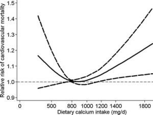
Calcium More or Less Than 800mg Increases Heart Risk, More or Less Than 900mg Increases All-Cause Mortality

Abstract

Dietary calcium intake and mortality risk from cardiovascular disease and all causes: a meta-analysis of prospective cohort studies.

Considerable controversy exists regarding the association between dietary calcium intake and risk of mortality from cardiovascular disease and all causes. Therefore, we performed a meta-analysis of prospective cohort studies to examine the controversy.
We identified relevant studies by searching MEDLINE, Embase, and the Cochrane Library databases between 1 September 2013 and 30 December 2013. Reference lists of relevant articles were also reviewed. Observational prospective studies that reported relative risks and 95% confidence intervals for the association of calcium intake with cardiovascular and all-cause mortality were eligible. Study-specific relative risks were pooled using a random-effects model.
In this meta-analysis, 11 prospective studies with 12 independent cohorts, involving 757,304 participants, were eligible. There was evidence of a non-linear association between dietary calcium intake and risk of mortality from cardiovascular disease (P for non-linearity <0.01) and all causes (P for non-linearity <0.01). A dose-response analysis showed a U-shaped relationship between dietary calcium intake and cardiovascular mortality. Intakes that were lower and higher than around 800 mg/day were gradually associated with a higher risk of cardiovascular mortality. For all-cause mortality, we also observed a threshold effect at intakes around 900 mg/day. The risk of all-cause mortality did not decrease further at intakes above 900 mg/day.
This meta-analysis of prospective cohort studies suggests that dietary calcium intake is associated with cardiovascular mortality in a U-shaped manner and that high dietary calcium intake (>900 mg/day) is not associated with a decreased risk of all-cause mortality.

Wang X, Chen H, Ouyang Y, Liu J…
BMC Med 2014
PMID: 25252963 | Free Full Text


From the full text:

Fig3

Calcium Supplements Associated with Increased Fracture Risk In Women

Abstract

Calcium intake and fracture risk: results from the study of osteoporotic fractures.

The relation between dietary calcium, calcium, and vitamin D supplements and the risk of fractures of the hip (n = 332), ankle (n = 210), proximal humerus (n = 241), wrist (n = 467), and vertebrae (n = 389) was investigated in a cohort study involving 9,704 US white women aged 65 years or older. Baseline assessments took place in 1986-1988 in four US metropolitan areas. Dietary calcium intake was assessed at baseline with a validated food frequency questionnaire. Data on new nonvertebral fractures were collected every 4 months during a mean of 6.6 years of follow-up; identification of new vertebral fractures was based on comparison of baseline and follow-up radiographs of the spine done a mean of 3.7 years apart. Results were adjusted for numerous potential confounders, including weight, physical activity, estrogen use, protein intake, and history of falls, osteoporosis, and fractures. There were no important associations between dietary calcium intake and the risk of any of the fractures studied. Current use of calcium supplements was associated with increased risk of hip (relative risk = 1.5, 95% confidence interval 1.1-2.0) and vertebral (relative risk = 1.4, 95% confidence interval 1.1-1.9) fractures; current use of Tums antacid tablets was associated with increased risk of fractures of the proximal humerus (relative risk = 1.7, 95% confidence interval 1.3-2.4). There was no evidence of a protective effect of vitamin D supplements. Although a true adverse effect of calcium supplements on fracture risk cannot be ruled out, it is more likely that our findings are due to inadequately controlled confounding by indications for use of supplements. In conclusion, this study did not find a substantial beneficial effect of calcium on fracture risk.

Cumming RG, Cummings SR, Nevitt MC, Scott J…
Am. J. Epidemiol. May 1997
PMID: 9149664 | Free Full Text