Tag Archives: free full text

Calcium Intake of 903mg to 1025mg Associated with Lowest Fracture Risk, More and Less Increases Risk

Abstract

Dietary calcium intake and risk of fracture and osteoporosis: prospective longitudinal cohort study.

To investigate associations between long term dietary intake of calcium and risk of fracture of any type, hip fractures, and osteoporosis.
A longitudinal and prospective cohort study, based on the Swedish Mammography Cohort, including a subcohort, the Swedish Mammography Cohort Clinical.
A population based cohort in Sweden established in 1987.
61,433 women (born between 1914 and 1948) were followed up for 19 years. 5022 of these women participated in the subcohort.
Primary outcome measures were incident fractures of any type and hip fractures, which were identified from registry data. Secondary outcome was osteoporosis diagnosed by dual energy x ray absorptiometry in the subcohort. Diet was assessed by repeated food frequency questionnaires.
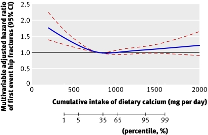
During follow-up, 14,738 women (24%) experienced a first fracture of any type and among them 3871 (6%) a first hip fracture. Of the 5022 women in the subcohort, 1012 (20%) were measured as osteoporotic. The risk patterns with dietary calcium were non-linear. The crude rate of a first fracture of any type was 17.2/1000 person years at risk in the lowest quintile of calcium intake, and 14.0/1000 person years at risk in the third quintile, corresponding to a multivariable adjusted hazard ratio of 1.18 (95% confidence interval 1.12 to 1.25). The hazard ratio for a first hip fracture was 1.29 (1.17 to 1.43) and the odds ratio for osteoporosis was 1.47 (1.09 to 2.00). With a low vitamin D intake, the rate of fracture in the first calcium quintile was more pronounced. The highest quintile of calcium intake did not further reduce the risk of fractures of any type, or of osteoporosis, but was associated with a higher rate of hip fracture, hazard ratio 1.19 (1.06 to 1.32).
Gradual increases in dietary calcium intake above the first quintile in our female population were not associated with further reductions in fracture risk or osteoporosis.

Warensjö E, Byberg L, Melhus H, Gedeborg R…
BMJ 2011
PMID: 21610048 | Free Full Text


From the full text:

• Dietary calcium intakes below approximately 700 mg per day in women were associated with an increased risk of hip fracture, any fracture, and of osteoporosis

• The highest reported calcium intake did not further reduce the risk of fractures of any type, or of osteoporosis, but was associated with a higher rate of hip fracture

fig2

Calcium Alone Increases Hip Fractures but Lowers Total Fractures

Abstract

Effect of calcium supplementation on hip fractures.

There have been numerous studies of the effects of calcium supplementation, with or without vitamin D, on fractures. Individually, they have not provided clarity regarding calcium’s anti-fracture efficacy, though they have established that calcium does have beneficial effects on bone density throughout the skeleton in women. Meta-analysis of these data suggests that total fracture numbers are diminished. However, the data from the 5,500 women involved in trials of calcium monotherapy show consistent adverse trends in numbers of hip fractures (relative risk 1.50, 95% CI 1.06-2.12). Observational data from the Study of Osteoporotic Fractures show a similar increase in risk of hip fracture associated with calcium use. We hypothesize that reduced periosteal expansion in women using calcium supplementation might account for the differences in anti-fracture efficacy of calcium at the hip, in comparison with other sites. Until there are further trial results to clarify this area, the present findings suggest that reliance on high calcium intakes to reduce the risk of hip fracture in older women is not appropriate. In addition, those at risk should be looking to other agents with a proven capacity to prevent hip fractures, such as bisphosphonates.

Reid IR, Bolland MJ, Grey A
Osteoporos Int Aug 2008
PMID: 18286218 | Free Full Text


Furthermore, our own recent trial of calcium monotherapy suggested that there might be heterogeneity between the responses of hip and other fractures to calcium supplementation [2], with downward trends in vertebral, forearm, and total osteoporotic fractures, but a significant increase in hip fractures.

[…]

Observational studies have also assessed the relationship between calcium use and fractures. While there is a potential problem of confounding by indication, it is noteworthy that the Study of Osteoporotic Fractures reported an increase in hip fracture risk in postmenopausal women taking calcium supplements of almost identical magnitude to that found in the present meta-analysis (relative risk 1.5; 95%CI, 1.1–2.0) [18]. This consistency across the available intervention studies and a large observational study raises doubts regarding the safety of calcium monotherapy in elderly postmenopausal women, though we cannot completely preclude the possibility that these results are a chance finding arising from the smaller numbers of this particular fracture type.

The adverse effect of calcium monotherapy on hip fractures poses the question of how this could occur when the same intervention has the opposite effect on total fracture numbers.

Resveratrol May Increase Bone Length in Pre-pubertal Rabbits

Abstract

Resveratrol treatment delays growth plate fusion and improves bone growth in female rabbits.

Trans-resveratrol (RES), naturally produced by many plants, has a structure similar to synthetic estrogen diethylstilbestrol, but any effect on bone growth has not yet been clarified. Pre-pubertal ovary-intact New Zealand white rabbits received daily oral administration of either vehicle (control) or RES (200 mg/kg) until growth plate fusion occurred. Bone growth and growth plate size were longitudinally monitored by X-ray imaging, while at the endpoint, bone length was assessed by a digital caliper. In addition, pubertal ovariectomized (OVX) rabbits were treated with vehicle, RES or estradiol cypionate (positive control) for 7 or 10 weeks and fetal rat metatarsal bones were cultured in vitro with RES (0.03 µM-50 µM) and followed for up to 19 days. In ovary-intact rabbits, sixteen-week treatment with RES increased tibiae and vertebrae bone growth and subsequently improved final length. In OVX rabbits, RES delayed fusion of the distal tibia, distal femur and proximal tibia epiphyses and femur length and vertebral bone growth increased when compared with controls. Histomorphometrical analysis showed that RES-treated OVX rabbits had a wider distal femur growth plate, enlarged resting zone, increased number/size of hypertrophic chondrocytes, increased height of the hypertrophic zone, and suppressed chondrocyte expression of VEGF and laminin. In cultured fetal rat metatarsal bones, RES stimulated growth at 0.3 µM while at higher concentrations (10 μM and 50 μM) growth was inhibited. We conclude that RES has the potential to improve longitudinal bone growth. The effect was associated with a delay of growth plate fusion resulting in increased final length. These effects were accompanied by a profound suppression of VEGF and laminin expression suggesting that impairment of growth plate vascularization might be an underlying mechanism.

Karimian E, Tamm C, Chagin AS, Samuelsson K…
PLoS ONE 2013
PMID: 23840780 | Free Full Text

Review: Orthosilicic Acid

Abstract

Biological and therapeutic effects of ortho-silicic acid and some ortho-silicic acid-releasing compounds: New perspectives for therapy.

Silicon (Si) is the most abundant element present in the Earth’s crust besides oxygen. However, the exact biological roles of silicon remain unknown. Moreover, the ortho-silicic acid (H4SiO4), as a major form of bioavailable silicon for both humans and animals, has not been given adequate attention so far. Silicon has already been associated with bone mineralization, collagen synthesis, skin, hair and nails health atherosclerosis, Alzheimer disease, immune system enhancement, and with some other disorders or pharmacological effects. Beside the ortho-silicic acid and its stabilized formulations such as choline chloride-stabilized ortho-silicic acid and sodium or potassium silicates (e.g. M2SiO3; M= Na,K), the most important sources that release ortho-silicic acid as a bioavailable form of silicon are: colloidal silicic acid (hydrated silica gel), silica gel (amorphous silicon dioxide), and zeolites. Although all these compounds are characterized by substantial water insolubility, they release small, but significant, equilibrium concentration of ortho-silicic acid (H4SiO4) in contact with water and physiological fluids. Even though certain pharmacological effects of these compounds might be attributed to specific structural characteristics that result in profound adsorption and absorption properties, they all exhibit similar pharmacological profiles readily comparable to ortho-silicic acid effects. The most unusual ortho-silicic acid-releasing agents are certain types of zeolites, a class of aluminosilicates with well described ion(cation)-exchange properties. Numerous biological activities of some types of zeolites documented so far might probably be attributable to the ortho-silicic acid-releasing property. In this review, we therefore discuss biological and potential therapeutic effects of ortho-silicic acid and ortho-silicic acid -releasing silicon compounds as its major natural sources.

Jurkić LM, Cepanec I, Pavelić SK, Pavelić K
Nutr Metab (Lond) 2013
PMID: 23298332 | Free Full Text


The full text article (link above) has a subsection on osteoporosis:

…Interestingly, the administration of silicon in a controlled clinical study induced a significant increase in femoral bone mineral density in osteoporotic women [31]. Direct relationship between silicon content and bone formation has been shown by Moukarzel et al. [64]. They found a correlation between decreased silicon concentrations in total parenterally fed infants with a decreased bone mineral content. This was the first observation of a possible dietary deficiency of silicon in humans. A randomized controlled animal study on aged ovariectomized rats revealed that long-term preventive treatment with ch-OSA prevented partial femoral bone loss and had a positive effect on the bone turnover [65]. Dietary silicon is associated with postmenopausal bone turnover and bone mineral density at the women’s age when the risk of osteoporosis increases. Moreover, in a cohort study on 3198 middle-aged woman (50–62 years) it was shown that silicon interacts with the oestrogen status on bone mineral density, suggesting that oestrogen status is important for the silicon metabolism in bone health [66].

Orthosilicic Acid Stimulates Bone Formation in Osteopenic Women

Abstract

Choline-stabilized orthosilicic acid supplementation as an adjunct to calcium/vitamin D3 stimulates markers of bone formation in osteopenic females: a randomized, placebo-controlled trial.

Mounting evidence supports a physiological role for silicon (Si) as orthosilicic acid (OSA, Si(OH)4) in bone formation. The effect of oral choline-stabilized orthosilicic acid (ch-OSA) on markers of bone turnover and bone mineral density (BMD) was investigated in a double-blind placebo-controlled trial.
Over 12-months, 136 women out of 184 randomized (T-score spine < -1.5) completed the study and received, daily, 1000 mg Ca and 20 microg cholecalciferol (Vit D3) and three different ch-OSA doses (3, 6 and 12 mg Si) or placebo. Bone formation markers in serum and urinary resorption markers were measured at baseline, and after 6 and 12 months. Femoral and lumbar BMD were measured at baseline and after 12 months by DEXA.
Overall, there was a trend for ch-OSA to confer some additional benefit to Ca and Vit D3 treatment, especially for markers of bone formation, but only the marker for type I collagen formation (PINP) was significant at 12 months for the 6 and 12 mg Si dose (vs. placebo) without a clear dose response effect. A trend for a dose-corresponding increase was observed in the bone resorption marker, collagen type I C-terminal telopeptide (CTX-I). Lumbar spine BMD did not change significantly. Post-hoc subgroup analysis (baseline T-score femur < -1) however was significant for the 6 mg dose at the femoral neck (T-test). There were no ch-OSA related adverse events observed and biochemical safety parameters remained within the normal range.
Combined therapy of ch-OSA and Ca/Vit D3 had a potential beneficial effect on bone collagen compared to Ca/Vit D3 alone which suggests that this treatment is of potential use in osteoporosis.

Spector TD, Calomme MR, Anderson SH, Clement G…
BMC Musculoskelet Disord 2008
PMID: 18547426 | Free Full Text


From the full text:

Collagen provides elasticity and structure in all connective tissues and several studies have indicated that collagen is important for bone toughness [43-45] whereas the mineral component is mainly involved in providing stiffness. Wang et al. [46] demonstrated that the mechanical integrity of collagen fibres deteriorates with ageing in human cortical bones and is associated with a higher fracture risk. When the collagen network becomes weaker with age, it will result in decreased toughness, possibly due to a reduction in natural cross-links or silicon content. It has previously been suggested that Si may be an integral (structural) component of connective tissues as high levels of non-dialysable Si has been reported in connective tissues and their components suggesting strong (covalent) associations [47].

Review: Silicon

Abstract

Silicon and bone health.

Low bone mass (osteoporosis) is a silent epidemic of the 21st century, which presently in the UK results in over 200,000 fractures annually at a cost of over one billion pounds. Figures are set to increase worldwide. Understanding the factors which affect bone metabolism is thus of primary importance in order to establish preventative measures or treatments for this condition. Nutrition is an important determinant of bone health, but the effects of the individual nutrients and minerals, other than calcium, is little understood. Accumulating evidence over the last 30 years strongly suggest that dietary silicon is beneficial to bone and connective tissue health and we recently reported strong positive associations between dietary Si intake and bone mineral density in US and UK cohorts. The exact biological role(s) of silicon in bone health is still not clear, although a number of possible mechanisms have been suggested, including the synthesis of collagen and/or its stabilization, and matrix mineralization. This review gives an overview of this naturally occurring dietary element, its metabolism and the evidence of its potential role in bone health.

Jugdaohsingh R
J Nutr Health Aging
PMID: 17435952 | Free Full Text

Strontium Ranelate Not Associated with Heart Attacks or Other Cardiac Events

Abstract

Ischaemic cardiac events and use of strontium ranelate in postmenopausal osteoporosis: a nested case-control study in the CPRD.

We explored the cardiac safety of the osteoporosis treatment strontium ranelate in the UK Clinical Practice Research Datalink. While known cardiovascular risk factors like obesity and smoking were associated with increased cardiac risk, use of strontium ranelate was not associated with any increase in myocardial infarction or cardiovascular death.
It has been suggested that strontium ranelate may increase risk for cardiac events in postmenopausal osteoporosis. We set out to explore the cardiac safety of strontium ranelate in the Clinical Practice Research Datalink (CPRD) and linked datasets.
We performed a nested case-control study. Primary outcomes were first definite myocardial infarction, hospitalisation with myocardial infarction, and cardiovascular death. Cases and matched controls were nested in a cohort of women treated for osteoporosis. The association with exposure to strontium ranelate was analysed by multivariate conditional logistic regression.
Of the 112,445 women with treated postmenopausal osteoporosis, 6,487 received strontium ranelate. Annual incidence rates for first definite myocardial infarction (1,352 cases), myocardial infarction with hospitalisation (1,465 cases), and cardiovascular death (3,619 cases) were 3.24, 6.13, and 14.66 per 1,000 patient-years, respectively. Obesity, smoking, and cardiovascular treatments were associated with significant increases in risk for cardiac events. Current or past use of strontium ranelate was not associated with increased risk for first definite myocardial infarction (odds ratio [OR] 1.05, 95 % confidence interval [CI] 0.68-1.61 and OR 1.12, 95 % CI 0.79-1.58, respectively), hospitalisation with myocardial infarction (OR 0.84, 95 % CI 0.54-1.30 and OR 1.17, 95 % CI 0.83-1.66), or cardiovascular death (OR 0.96, 95 % CI 0.76-1.21 and OR 1.16, 95 % CI 0.94-1.43) versus patients who had never used strontium ranelate.
Analysis in the CPRD did not find evidence for a higher risk for cardiac events associated with the use of strontium ranelate in postmenopausal osteoporosis.

Cooper C, Fox KM, Borer JS
Osteoporos Int Feb 2014
PMID: 24322476 | Free Full Text

Depression Associated With Bone Mass in Premenopausal Women

Abstract

Do premenopausal women with major depression have low bone mineral density? A 36-month prospective study.

An inverse relationship between major depressive disorder (MDD) and bone mineral density (BMD) has been suggested, but prospective evaluation in premenopausal women is lacking.
Participants of this prospective study were 21 to 45 year-old premenopausal women with MDD (n = 92) and healthy controls (n = 44). We measured BMD at the anteroposterior lumbar spine, femoral neck, total hip, mid-distal radius, trochanter, and Ward’s triangle, as well as serum intact parathyroid hormone (iPTH), ionized calcium, plasma adrenocorticotropic hormone (ACTH), serum cortisol, and 24-hour urinary-free cortisol levels at 0, 6, 12, 24, and 36 months. 25-hydroxyvitamin D was measured at baseline.
At baseline, BMD tended to be lower in women with MDD compared to controls and BMD remained stable over time in both groups. At baseline, 6, 12, and 24 months intact PTH levels were significantly higher in women with MDD vs. controls. At baseline, ionized calcium and 25-hydroxyvitamin D levels were significantly lower in women with MDD compared to controls. At baseline and 12 months, bone-specific alkaline phosphatase, a marker of bone formation, was significantly higher in women with MDD vs. controls. Plasma ACTH was also higher in women with MDD at baseline and 6 months. Serum osteocalcin, urinary N-telopeptide, serum cortisol, and urinary free cortisol levels were not different between the two groups throughout the study.
Women with MDD tended to have lower BMD than controls over time. Larger and longer studies are necessary to extend these observations with the possibility of prophylactic therapy for osteoporosis.
ClinicalTrials.gov NCT 00006180.

Cizza G, Mistry S, Nguyen VT, Eskandari F…
PLoS ONE 2012
PMID: 22848407 | Free Full Text

Review: Calcium Safety and New Recommendations

Abstract

Calcium builds strong bones, and more is better–correct? Well, maybe not.

Calcium supplementation has been considered the gold standard therapy for osteoporosis in the general population. It is given in both the placebo and treatment groups of trials evaluating antifracture efficacy of new therapies. Similarly, calcium-based phosphate binders have been considered the gold standard comparator for all new phosphate binders. However, large randomized trials demonstrate conflicting data on the antifracture efficacy of calcium supplementation, particularly in high doses, in patients with osteoporosis without CKD. In addition, recent data suggest an increased risk for cardiovascular events. These new studies raise safety concerns for the general approach with calcium supplementation and binders. This review describes recent data on the adverse effects of calcium supplementation for osteoporosis and how these new data should affect the strategy for phosphate binder use in CKD.

Jamal SA, Moe SM
Clin J Am Soc Nephrol Nov 2012
PMID: 22837272 | Free Full Text


It is important to note that some clinical practice guidelines have been modified on the basis of this new literature suggesting potential risk. For example, in its recently published evidence-based guidelines, Osteoporosis Canada recommended a total intake of calcium (from diet and supplement) of 1200 mg per day, a decrease from the previous recommendation of 1500 mg in supplements (32). The American Society for Bone and Mineral Research issued a statement regarding the potential risks of calcium supplements and suggested, among other points, that “the beneficial effects of calcium are found with relatively low doses. More is not necessarily better. Individuals should discuss the amount of their calcium intake with their healthcare provider” (33). The Institute of Medicine now recommends a daily dietary reference allowance of calcium of 1000–1200 mg per day in the form of diet and supplements (34,35). Finally, the draft United States Preventive Services Task Force statement, pending public comment (http://www.uspreventiveservicestaskforce.org/draftrec3.htm), currently states “the current evidence is insufficient to assess the balance of the benefits and harms of combined vitamin D and calcium supplementation for the primary prevention of fractures in premenopausal women or in men.” Thus, these authorities acknowledge that although some calcium supplements may be beneficial for bone health, too much calcium may be harmful.

Review: Incretins and Bone

Abstract

Incretins and bone: evolving concepts in nutrient-dependent regulation of bone turnover.

Postprandial variation of bone turnover markers and the closed relationship between bone remodeling and nutrient supply has been extensively studied in the past few years, but the underlying pathophysiologic mechanisms remain largely unknown. Recent studies have shown that the acute regulation of bone turnover induced by feeding is probably mediated by gastrointestinal (GI) peptides. The greater response of bone remodeling during oral versus intravenous glucose administration and the inhibition of this response after administration of octreotide, that inhibits the release of GI peptides, further support the existence of a gutbone axis. Glucose-dependent insulinotropic peptide and glucagon-like peptides-1 and -2 are released from K and L cells of the gastrointestinal tract, respectively, and are considered the main mediators of the postprandial response of bone turnover. In this review we outline the most recent evidence that demonstrates the role of incretins in nutrient-dependent regulation of bone metabolism. Further elucidation of the underlying mechanisms can be exploited therapeutically in the future.

Yavropoulou MP, Yovos JG
Hormones (Athens)
PMID: 23933690 | Free Full Text